
Medical imaging technology has transformed healthcare by providing non-invasive methods to visualise internal structures, facilitating diagnosis, treatment planning, and disease monitoring. Within the realm of medical imaging, women’s imaging represents a specialised field that focuses on addressing the unique healthcare needs of women. In this article, we explore the diverse clinical applications of imaging technology, explicitly emphasising women’s imaging and its crucial role in improving women’s health outcomes.

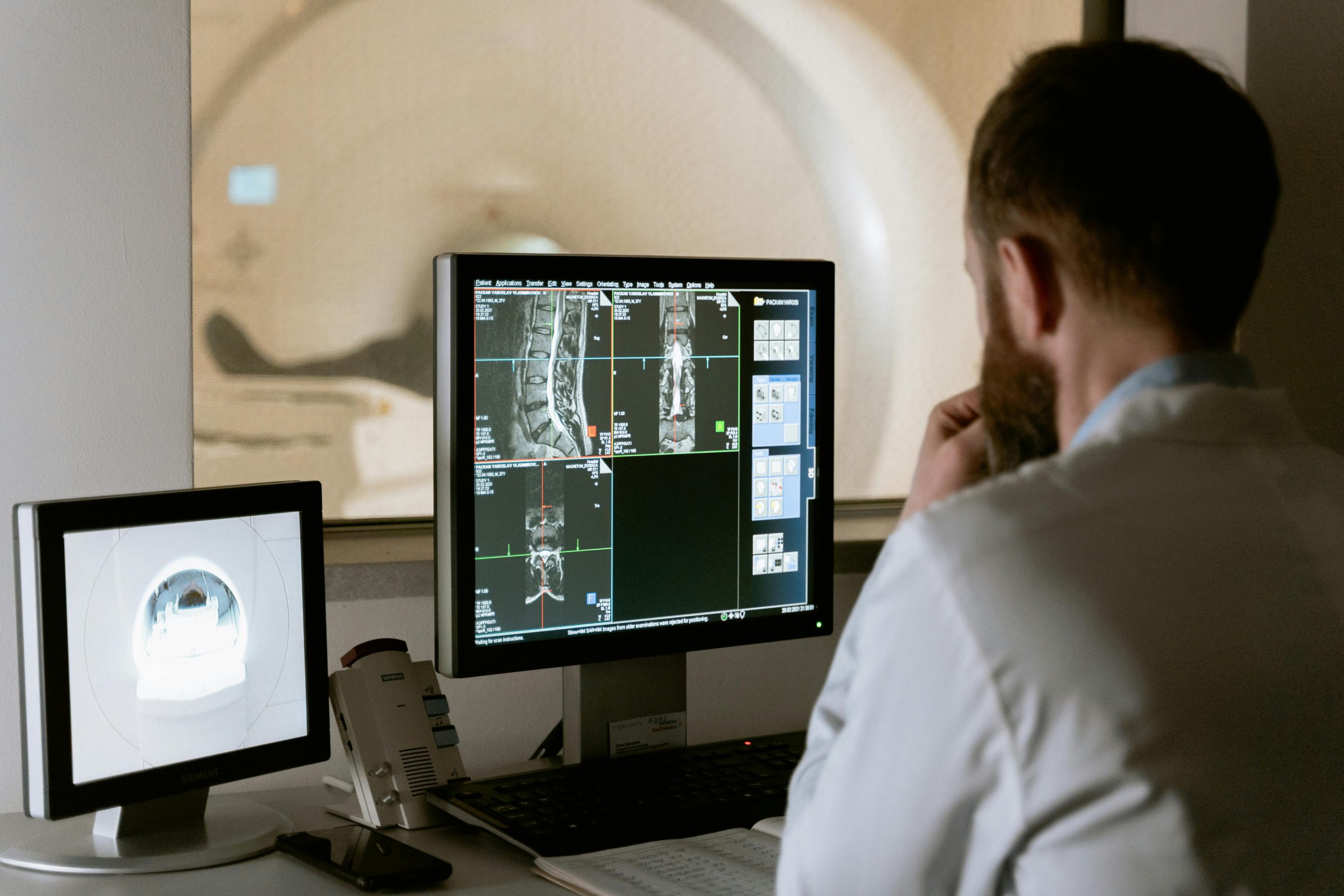
- Diagnostic Imaging: One of imaging technology’s fundamental applications is diagnosis. Medical imaging modalities such as X-rays, computed tomography (CT), magnetic resonance imaging (MRI), and ultrasound are employed to visualise anatomical structures and detect abnormalities. In women’s imaging, these modalities play a vital role in diagnosing conditions specific to women, such as breast cancer, ovarian cysts, uterine fibroids, and pelvic inflammatory disease.
- Breast Imaging: Breast imaging encompasses a range of modalities used to detect and evaluate breast conditions. Mammography is the cornerstone of breast cancer screening, enabling the early detection of abnormalities such as tumours or calcifications. Additionally, breast ultrasound and MRI are valuable adjuncts for further characterization of breast lesions and guiding biopsy procedures. These imaging techniques are essential components of women’s healthcare, aiding in the early detection and management of breast cancer and other breast-related conditions.
- Obstetric Imaging: Obstetric imaging is vital in monitoring foetal development and ensuring maternal-foetal health during pregnancy. Ultrasound is the primary imaging modality used for obstetrics, allowing for visualising the foetus, placenta, and maternal reproductive organs. Obstetric ultrasound assesses foetal growth, detects congenital anomalies, and monitors maternal conditions such as placenta previa or uterine abnormalities. Advanced techniques such as 3D/4D ultrasound provide detailed anatomical information and enhance the prenatal bonding experience for expectant parents.
- Gynecologic Imaging: Gynecologic imaging evaluates the female reproductive system and related conditions. Transvaginal ultrasound is commonly used to assess the uterus, ovaries, and fallopian tubes, aiding in the diagnosis of conditions such as ovarian cysts, endometriosis, and uterine fibroids. MRI is also employed in gynecologic imaging to provide additional information about pelvic anatomy and pathology, particularly in cases where ultrasound findings are inconclusive or further characterization is needed.
- Bone Density Imaging: Bone density imaging, also known as dual-energy X-ray absorptiometry (DEXA), assesses bone mineral density and diagnoses osteoporosis. DEXA scans are commonly performed in women to evaluate fracture risk and monitor response to osteoporosis treatment. Early detection and intervention are crucial in preventing osteoporotic fractures, particularly in postmenopausal women who are at increased risk due to hormonal changes.
- Fertility Imaging: Fertility imaging encompasses imaging techniques used to assess reproductive function and aid in managing infertility. Transvaginal ultrasound is commonly used to evaluate ovarian reserve, assess the uterine cavity for abnormalities, and monitor follicular development during ovulation induction. Hysterosalpingography (HSG) is another imaging modality to assess the fallopian tubes and uterine cavity for obstruction or structural abnormalities that may impact fertility.
- Interventional Imaging: Interventional imaging involves using imaging guidance to perform minimally invasive procedures for diagnostic or therapeutic purposes. In women’s imaging, interventional procedures may include image-guided breast biopsy, ultrasound-guided ovarian cyst aspiration, or hysterosalpingography with tubal catheterization for fertility evaluation. These procedures offer the advantage of targeted tissue sampling or treatment delivery while minimising patient discomfort and recovery time.
- Screening and Preventive Imaging: Screening and preventive imaging play a crucial role in women’s healthcare by enabling early detection and intervention for asymptomatic conditions. Mammography screening programs have been instrumental in reducing breast cancer mortality by detecting tumours at an earlier, more treatable stage. Additionally, imaging techniques such as pelvic ultrasound or MRI may be used for screening or surveillance in women at increased risk for gynecologic malignancies or other reproductive health conditions.
Imaging technology encompasses a wide range of clinical applications integral to women’s healthcare. From diagnostic imaging for breast cancer screening to obstetric ultrasound for monitoring foetal development, imaging modalities play a crucial role in the prevention, diagnosis, and treatment of various women’s health conditions. By leveraging the capabilities of imaging technology, healthcare providers can deliver personalised, effective care that meets the unique needs of women throughout their lifespan.